Author: Dr Tho Kam San
Date: May 2011 Issue
Original Blog Link: Link
As discussions about the hot seat in parliament continue, it’s hard to believe we’ve already reached the middle of 2011. Now in its third edition, Ortho Insider focuses on various ligament injuries and how our orthopaedic doctors address them based on their experience.
When managing knee ligament injuries, the primary goal is to restore normal knee movement, stability, and function. Commonly, single ligament injuries like sprains to the medial collateral ligament (MCL) or anterior cruciate ligament (ACL) occur during twisting movements.
MCL Injuries:
Isolated MCL injuries, grade I to II, are treated conservatively with knee braces, analgesics and physiotherapy. These injuries recover in 4-6 weeks; in professional soccer players light physical exercises are then started. Grade III injuries often have significant swelling and bruising medially with valgus stress opening >1cm. MRI will show a complete tear. These injuries require surgical repair. Post surgery, the knee is braced in full extension for 4 weeks with a gradual increase in flexion in the brace for next 2 weeks. Generally, full function is regained 3 months post surgery.
ACL Injuries:
Isolated ACL tears occur with twisting injuries to the knee, most commonly during soccer. Minor sprains will recover after 4-6 weeks with rest and physiotherapy. Partial tears often recover with conservative treatment. In cases of instability, the knee should be assessed for associated ligament injuries. Surgery should be considered for persistent instability despite intensive therapy.
In complete ACL tears, it is important to exclude associated ligament injuries. Isolated ACL tears in physically active adults often require surgery. My experience with professional soccer players suggest the need for surgery if one plans to continue with sports which require pivoting.
In patients who stop pivoting sports, physiotherapy may suffice to stabilise the knee. However, instability during activities of daily living will require surgery as frequent giving way of the knee will damage the cartilage and meniscus leading to early onset of osteoarthritis. Hence, ACL reconstruction (ACLR) is often a need and lifestyle choice.
ACLR is a common reconstructive procedure which can be done as a day surgery procedure. The graft is based on the hamstring tendons (semitendinosus, gracillis), or patella bone-tendon-bone (PBTB) graft or rarely, the quadriceps tendon. I prefer the semitendinosus tendon folded twice to give a thick 4 strand graft. The gracilis is harvested only if the semitendinosus is not long enough. PBTB grafts are strong but donor site problems like patellofemoral pain can be quite debilitating. I usually reserve PBTB grafts for revision cases. If that ruptures, the quadriceps tendon is next. Allograft can be used, but the strength is not ideal, especially lyophilised grafts. Fresh frozen grafts are better in quality but there is the risk of cross infection, although highly remote. Allografts are indicated in multiple ligament reconstructions where graft availability is a problem.
Double tunnel ACLR is a recent development with many variations that may offer better rotational stability by reconstructing a more anatomical ACL graft. Dividing the graft into two separate smaller bundles however compromises graft strength. Drilling one set of tunnels precisely can be challenging; drilling 2 sets of tunnels accurately, even more so. Double Tunnel = Double Trouble. Current long term results of single bundle ACLR are excellent with over 90% success rate. Until the ideal patient and the ideal double tunnel method are defined, single bundle ACLR will remain the technique of choice. If there is associated posteromedial corner or postero-lateral corner (PLC) instability, extra-articular reconstruction is performed using the ilio-tibial band or allograft.
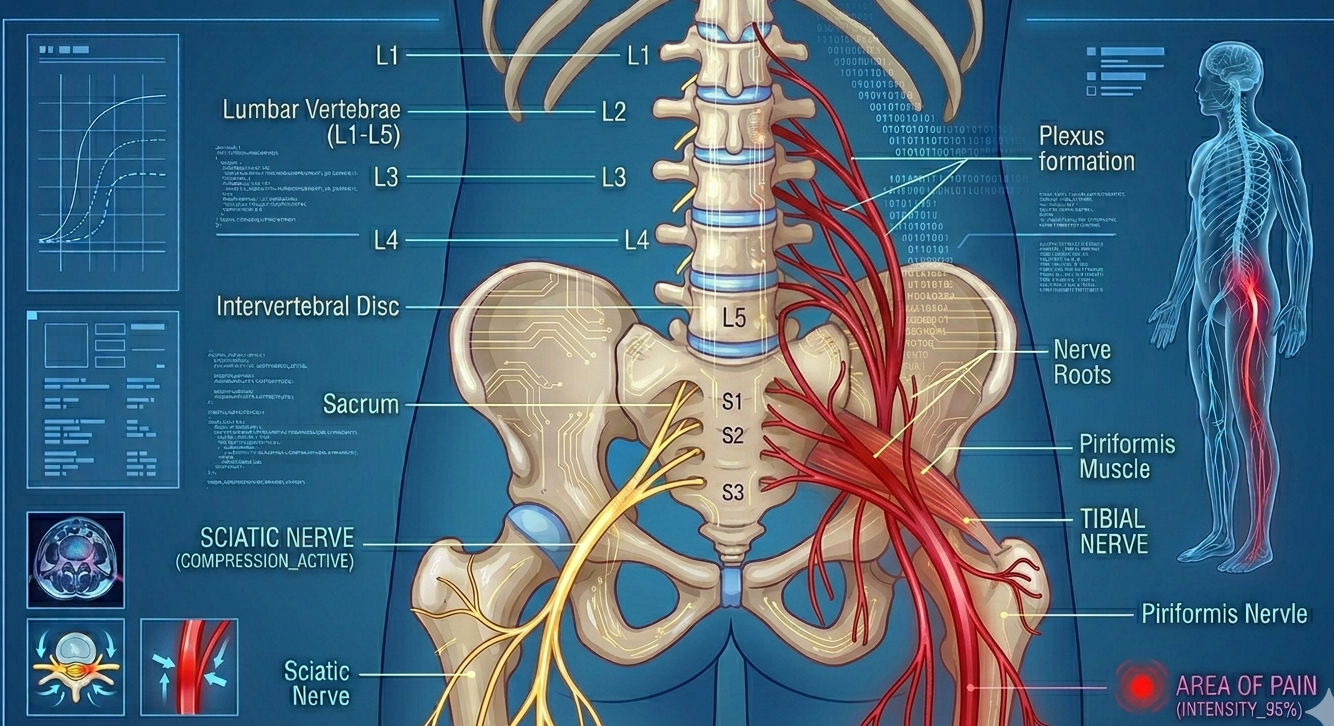
Posterior Cruciate Ligament (PCL):
PCL injuries are uncommon, often from a direct anterior pretibial force like a dash-board injury or a hyper flexion injury. Classically, there is a posterior sag. MRI is confirmatory. PCL tears are often less debilitating than ACL tears. Hence, early bracing and physiotherapy often suffice. Arthroscopic PCLR (with hamstring or PBTB) is performed only for unstable knees. In cases of a PCL bony avulsion injury where the bone fragment is displaced by more than 1cm, surgical re-attachment advised. This can be done arthroscopically. In chronic PCL tears with instability, PLC insufficiency is not uncommon. In these cases, the PLC also requires reconstruction.
Multi-Ligament Injuries:
Multiple knee ligament injuries are seen in severe soccer injuries, vehicular accidents and more recently, skiing accidents. The common combinations are ACL & PCL +/- MCL. In these severe injuries, popliteal artery and nerve damage must be excluded. It is best to reconstruct these injuries early before massive swelling sets in. Otherwise, the knee is braced and the swelling allowed to settle prior to surgery. I routinely use PBTB for the ACL, hamstring for the PCL and repair the MCL with the aid of suture anchors. Post surgery physiotherapy is critical for a successful outcome. Physiotherapy is essential to help regain optimal knee function. With timely surgical intervention and intensive physiotherapy, most patients will return to their pre-injury level of physical activity.
More about the Author:
Dr. Tho Kam San is the team physician to the National Soccer Team where treatment for ligament injuries is the biggest part of the work. This contributes to his rich experience, many of them are penned down in publications in international and local journals.
LARGEST-EVER BIOPSY STUDY IN OSTEOPOROSIS – RESULTS COMMUNICATION ECCEO 2011 – Valencia, 24 March 2011
Bone biopsy is the gold standard technique used to examine the effect of osteoporosis treatments on bone. Bone biopsies involve taking a cylindrical sample of real bone from the upper part of the pelvis called the iliac crest. Biopsies allow the identification of non-mineralized (osteoid tissue) and mineralized subparts of the bone matrix and can be used to measure bone formation rates and other parameters. Bone formation is measured by examining mineralizing surface, the proportion of bone surface on which new mineralized bone is being deposited.
In this international, double-blind study of 268 women, Strontium Ranelate has a significantly greater effect on mineralizing surface compared to alendronate. After six months, mineralizing surface, expressed as a percentage of bone surface (the study’s primary endpoint) was 2.94% in patients receiving Strontium Ranelate compared to 0.20% in patients receiving alendronate (p<0.001). This superior effect on bone formation was further amplified after 12 months of treatment. Strontium Ranelate also significantly increased both the bone formation rate, and the mineral apposition rate compared to alendronate over six and 12 months of treatment. The superior bone-forming efficacy of Strontium Ranelate is linked to its innovative dual mechanism of action which rebalances bone turnover in favor of the formation of newer and stronger bone. This is not the case with bisphosphonates as they have been shown to suppress the bone forming surface.
“The results of the study show that Strontium Ranelate preserves a higher bone forming activity compared with alendronate”, notes study investigator Professor Roland Chapurlat from the Hôpital Edouard Herriot, Service de Rhumatologie et Pathologie Osseuse, Lyon, France.
“The bone-forming activity seen here can be attributed to strontium ranelate’s unique mechanism of action which, unlike bisphosphonates that block bone resorption and formation, combines the dual effects of increasing or maintaining bone formation and decreasing bone resorption”, points out study investigator Professor PG Ste- Marie from the Centre Hospitalier de l’Université de Montréal, Canada. This new trial conducted in 268 post-menopausal women with osteoporosis is the largest biopsy study ever. It sets a new standard in the assessment of the effects of different osteoporosis treatments on bone.
References
Chavassieux P, Brixen K, Zerbini C, et al. Bone formation is significantly greater in women on strontium ranelate than in those on alendronate after 6 and 12 months of treatment: histomorphometric analysis from a large randomized controlled trial. Osteoporos Int. 2011. Abstract OC16. Presented at the ECCEO Congress, Valencia, Spain, March 2011. Ott SM. Long-term safety of bisphosphonates. J Clin Endocrinol Metab 2005; 90:1897-9